Commotion cérébrale d'origine sportive
Il est maintenant bien connu qu'une commotion cérébrale chez l'adulte cause des perturbations cognitives importantes qui peuvent persister jusqu'à deux ans après l'incident. Cependant, même si l'incidence d'une commotion cérébrale est aussi élevée chez l'enfant que chez l'adulte, nos connaissances des séquelles cognitives et sensorielles chez l'enfant commotionné sont très limitées. Les objectifs de ce volet de recherche sont les suivants : 1) déterminer la nature des déficits neuropsychologiques causés par une commotion durant le développement ; 2) identifier les dysfonctions neurophysiologiques associées ; 3) déterminer s'il y a une relation entre l'âge auquel la commotion est survenue et la gravité des déficits ; 4) identifier les mécanismes de récupération et les limites de la plasticité des différentes habiletés cognitives et sensorielles en fonction de l'âge ; et 5) développer en dernier lieu un outil diagnostique pour les enfants commotionnés qui tiendra compte de l'âge auquel l'accident est survenu. Cet axe de recherche vise également à identifier les conséquences des commotions cérébrales sur les plans neurophysiologique et neurochimique et à déterminer si les séquelles des commotions cérébrales diffèrent entre les hommes et les femmes.
Projets récents :
Neuropsychological and neurophysiological deficits following a sport-related concussion in children and adolescents
We determined whether age differences exist with respect to cognitive functioning following a sport-related concussion. To do so, 96 athletes were recruited [9-12 yrs (n = 32); 13-16 yrs (n =34); adults (n = 30)] half of whom suffered from a sport-related concussion. Cognitive functioning has been assessed using standardized neuropsychological tests as well as event-related potentials elicited by a visual 3-stimulus oddball paradigm. The PCSS was used to assess symptoms experienced at the time of injury. Neuropsychological assessment with an adaptation of the testing battery used by the National Hockey League. Latencies and amplitudes of the P3a and P3b ERP components were analysed in terms of group (concussed vs control) and age. Children and adults did not present neuropsychological deficits 6 months after their last concussion (ps > .05). In contrast, adolescents showed persistent deficits in working memory (p ˂ .05). Concussed athletes displayed significantly lower amplitude for the P3b component of their ERP compared to their non injured teammates (p ˂ .05). No age-related differences were found among the concussed groups (p > 0.05). These data suggest persistent neurophysiological deficits that are present at least 6 months following a concussion. Moreover, the present findings suggest that adolescents are more sensitive to the consequences of concussions than are children or adults.
Auditory processing disorder following a sport-related concussion
The aim of the study is to investigate whether sport-related concussions disrupt auditory processes. Sixteen university athletes participated in the study: eight had one or more sport-related concussions and eight never experienced a concussion. The Frequency Pattern Sequence test, the Duration Pattern Sequence test, the Synthetic Sentence Identification test, and the Staggered Spondaic Word test were used to assess auditory processing. All nonconcussed athletes have normal auditory processing. In contrast, more than half of the concussed athletes had deficits for one or more of the auditory processing tests. The pattern of results suggests that sport-related concussions can disrupt the neurological mechanisms implicated in several auditory processes, including monaural low-redundancy speech recognition, tone pattern recognition, and dichotic listening.
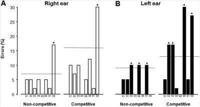
Performances of concussed (S1–S8) athletes (%
errors) on the four test conditions (right noncompeting;
right competing; left competing; left noncompeting) of
the Staggered Spondaic Word test. The dashed line
represents 2 SD above the mean of the control group.
Asterisk (*) represents scores 2 SD above the control
group and identify participants with abnormal results.
The white bars represent datafrom the right ear and the
black bars represent data from the left ear.
Prolonged neuropsychological impairments following a first concussion in female university soccer athletes
Although research is accumulating on the cognitive sequelae from sports-related concussions in men, little to nothing is known about the prolonged cognitive outcome following a concussion in women. This point is important because recent evidence suggests that female athletes are at greater risk of sustaining a concussion. We assessed cognitive functioning following a first concussion in female soccer players, 6 to 8 months after their injury. The first-time concussed athletes were compared to a group of age-matched team-mates who never experienced a concussion. A total of 22 female university-level soccer players participated in the study. Paper-pencil and computerized tasks were used to assess different neuropsychological functions. Short- and long-term verbal memory, attention, and simple reaction-time were normal. In contrast, compared to the control group the concussed athletes were significantly slower on tasks that required decision making (complex reaction-time), inhibition and flexibility (Stroop), and planning (Tour of London task). The results of this study suggest that cognitive functions related to cognitive processing speed are most vulnerable to a sports-related concussion and are still impaired over half a year post-injury in university-level female soccer players.
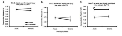
Metabolic changes in the acute and chronic post-concussion phases
Despite negative neuroimaging findings many athletes display neurophysiological alterations and post-concussion symptoms that may be attributable to neurometabolic alterations. The present study investigated the effects of sports concussion on brain metabolism using 1H-MR Spectroscopy by comparing a group of 10 non-concussed athletes with a group of 10 concussed athletes of the same age (mean: 22.5 years) and education (mean: 16 years) within both the acute and chronic post-injury phases. All athletes were scanned 1-6 days post-concussion and again 6-months later in a 3T Siemens MRI. Concussed athletes demonstrated neurometabolic impairment in prefrontal and motor (M1) cortices in the acute phase where NAA:Cr levels remained depressed relative to controls. There was some recovery observed in the chronic phase where Glu:Cr levels returned to those of control athletes; however, there was a pathological increase of m-I:Cr levels in M1 that was only present in the chronic phase. These results confirm cortical neurometabolic changes in the acute post-concussion phase as well as recovery and continued metabolic abnormalities in the chronic phase. The results indicate that complex pathophysiological processes differ depending on the post-injury phase and the neurometabolite in question.